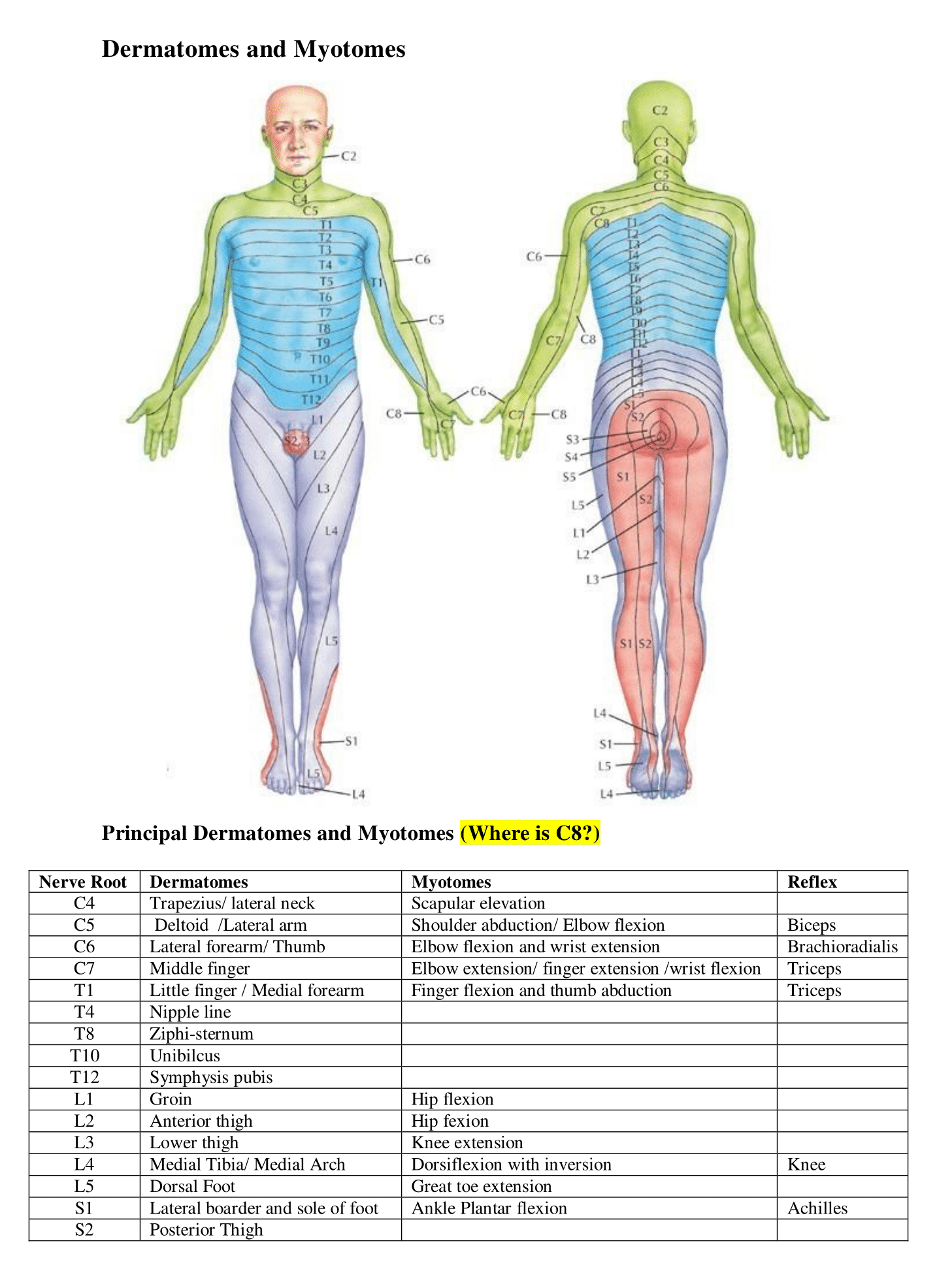
Useful Anatomy (https://galwayem.ie/index.php/resources/useful-anatomy)
Normal Radiographic Anatomy of the Foot
Image

- Medial malleolus
- Lateral malleolus
- Head of talus
- Calcaneus
- Navicular
- Cuboid
- Base of fifth metatarsal
- Third cuneiform (lateral)
- Second cuneiform (Intermediate)
- First cuneiform (medial)
- Medial sesamoid
- Lateral sesamoid
- Shaft of second metatarsal
- Neck of third metatarsal
- Head of fourth metatarsal
- Metatarsophalangeal joint
- Proximal phalanx
- Middle phalanx
- Distal phalanx
Case courtesy of Dr Benoudina Samir, Radiopaedia.org (http://radiopegia.org).
From the case rID: 49119 (http://Case courtesy of Dr Benoudina Samir, Radiopaedia.org. From the case rID: 49119)Normal radiographic anatomy of the wrist
Image

Image

- metacarpal bone
- carpometacarpal joint
- trapezoid
- trapezium
- scapho-trapezio-trapezoid (STT) joint
- scaphoid
- styloid process of radius
- radiocarpal joint
- lunate
- radius
- ulna
- distal radioulnar joint
- styloid process of ulna
- triquetrum
- pisiform
- capitate
- hamate
- hook of the hamate
Case courtesy of Dr Benoudina Samir,Radiopaedia.org (https://radiopaedia.org).
From the case rID: 43305 (https://radiopaedia.org/cases/43305)